TANGENTIAL INTERTUBERCULAR SULCUS
Specialized projection for evaluation of the humeral intertubercular sulcus
Demonstrated Pathology
- Pathologies of the intertubercular sulcus
- Evaluation of the biceps brachii tendon
- Fractures of the intertubercular sulcus
- Osteoporosis and osteoarthritis
- Degenerative changes of the sulcus
Exposure Factors
Low exposure: Parameters optimized for tangential visualization of the sulcus
Visible Anatomical Structures
The following should be clearly observed:
- Intertubercular sulcus (bicipital groove) in profile
- Greater and lesser tuberosities of the humerus
- Proximal portion of the humerus
- Relationship of the biceps tendon with the sulcus
- Thickness and contours of the sulcus
Plate Size
Positioning Variations
Standing Position
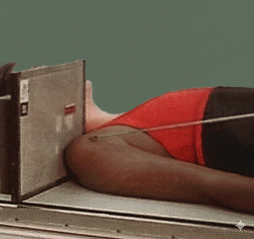
Supine Position
Central Ray by Position
Standing Position
Direction: Perpendicular
Point: Sulcus area at mid-anterior margin of humeral head
Supine Position
Direction: 10-15° behind horizontal
Point: Sulcus at mid-anterior margin of humeral head
Instructions to the Patient
"Hold your breath during exposure"
Maintain leaning position and chassis support without moving
Optimal Image Characteristics
Visible Sulcus
Bicipital groove in profile
No Superimposition
Differentiated bony structures
Adequate Depth
Sulcus without distortion
Centered Field
Proximal portion of the humerus
Common Technical Challenges
Frequent problems in tangential projection:
- Incorrect inclination of the humerus (not 10-15°)
- Poor chassis placement relative to the shoulder
- Wrong central ray angulation
- Patient movement due to uncomfortable position
- Superimposition of structures due to inadequate rotation
Solution: Ensure 10-15° inclination of the humerus and correct CR angulation according to position